Cytology vs. Histopathology
October 18, 2023
Dogs and cats grow new lumps or bumps pretty commonly – what are we supposed to do about them? It’s absolutely reasonable to ask your veterinarian to take a peek and make recommendations.
Sometimes, the new bumps are nothing more than a bug bite, a skin infection, or something minor that we don’t worry about too much. Other times, your veterinarian may recommend follow-up tests: cytology (via fine needle aspiration, skin scrape, impression smear, or swabbing) and/or histopathology (via biopsy).
What’s the difference?! And why is biopsy so much more expensive and involved?
| Cytology | Histopathology | |
|---|---|---|
| How? | Impression Smear: we use tape or a slide to get some cells off the surface of the skin and examine them under the microscope. Fine needle aspirate: inserting a small needle into the mass to draw out a few cells to examine under the microscope. | Remove a small piece of whole tissue via punch biopsy, excisional biopsy (surgically moving the entire mass), or incisional biopsy (surgically removing part of the mass). |
| What are we looking at? | Just a few cells – this is sometimes diagnostic, unfortunately sometimes it’s not | An entire piece of tissue aka “tissue architecture” – if the sample is appropriately taken, this almost always provides a definitive diagnosis |
| Where in the process? | Often the first step, can usually be performed on an awake patient | Usually not the first step, sedation or general anesthesia required |
| Who samples the mass? | Your primary care veterinarian | Your primary care veterinarian (who sends it off to a pathologist) |
| Who can perform the test and provide results? | Your primary care veterinarian or a veterinary pathologist | Your regular veterinarian can obtain the sample, but they submit the tissue to a veterinary pathologist for evaluation |
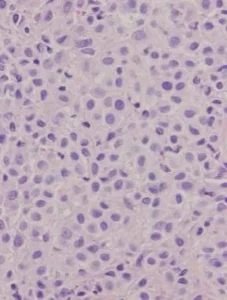
Cytology
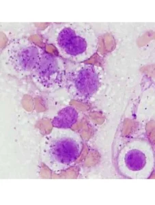
Histopathology
Written by Dr. Lauren Tanner
Related Posts
DVM Blog Post - Osteoarthritis AKA Degenerative Joint Disease
CAC Staff | September 12, 2024
Osteoarthritis, also known as degenerative joint disease, is a condition which will affect most dogs in their lifetime. As dogs age the incidence of arthritis increases and other confounding...